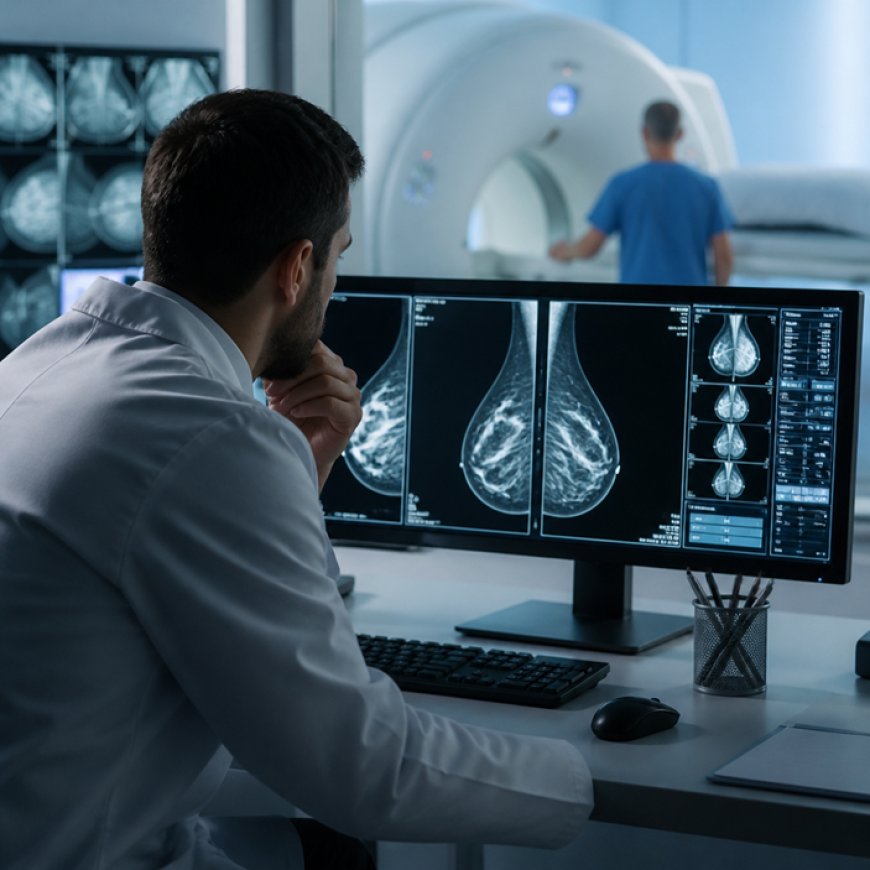
The conversation around breast cancer screening is shifting from a one-size-fits-all approach to risk-stratified protocols that match imaging intensity to individual patient profiles. Women with dense breasts, genetic predispositions, or prior atypical findings are not well-served by annual mammograms alone. Recent discussions in oncology journals emphasise the need for supplemental imaging in these populations, acknowledging that early detection in high-risk groups demands more sensitive tools.
The Dense Breast Dilemma
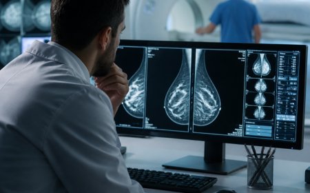
Dense breast tissue is not a pathology but a common anatomical variation. On a mammogram, both glandular tissue and tumours appear white, creating a visual challenge akin to spotting a snowflake in a snowstorm. Studies estimate that mammography's sensitivity drops from around ninety per cent in fatty breasts to as low as sixty per cent in extremely dense tissue. This means up to four in ten cancers could be missed in women with dense breasts if mammography is the sole screening method.
Legislation in several countries now requires radiologists to inform patients of breast density status, but notification alone does not solve the problem. Women are left navigating fragmented guidance on what to do next. Some are offered ultrasound as an adjunct, which improves detection but increases false positives and callback rates. Others receive no follow-up recommendation at all, creating a knowledge gap that can delay diagnosis by months or years.
How Advanced Imaging Closes the Gap
Supplemental imaging technologies offer a solution by using contrast, magnetic fields, or three-dimensional reconstruction to differentiate tissue types with greater precision. Contrast-enhanced spectral mammography injects a contrast agent that highlights areas of increased blood flow, a hallmark of malignancy. Automated breast ultrasound scans the entire breast in thin slices, revealing masses that might be obscured on two-dimensional images. Molecular breast imaging uses radioactive tracers to detect metabolic activity in tumours, providing functional rather than purely anatomical information.
Each modality carries trade-offs. Contrast-enhanced techniques require intravenous access and carry a small risk of allergic reaction. Molecular imaging involves low-dose radiation exposure. Ultrasound is operator-dependent and time-intensive. Yet in women with dense breasts or elevated genetic risk, these methods consistently detect cancers missed by mammography, often at earlier stages when treatment is less aggressive and more effective.
MRI as a High-Sensitivity Standard
For women at significantly elevated risk—those with BRCA mutations, strong family histories, or prior chest radiation—guidelines have long endorsed annual screening with both mammography and MRI. The latter has emerged as the most sensitive imaging tool available, capable of detecting cancers smaller than five millimetres and distinguishing between benign and malignant lesions based on enhancement patterns. Facilities offering comprehensive breast health services, particularly in regions with robust healthcare infrastructure, increasingly integrate magnetic resonance imaging into screening protocols for high-risk populations, often alongside genetic counselling and advanced diagnostic services such as image-guided biopsy and reconstructive planning.
The sensitivity of MRI comes with a corresponding increase in false positives. Approximately ten to fifteen per cent of screening MRIs result in callbacks, many of which lead to benign findings on biopsy. This higher callback rate is the price of catching more cancers early, and for women at elevated risk, the trade-off is generally considered worthwhile. The key is appropriate patient selection: MRI is not recommended for average-risk women with non-dense breasts, where the false-positive burden would outweigh the benefit.
Integrating Imaging into Multidisciplinary Care
Detection is only the first step. Once a suspicious finding emerges, patients enter a cascade of diagnostic procedures—additional imaging, biopsy, pathology review—that requires coordination across specialties. The most effective breast cancer programmes embed radiologists, surgeons, medical oncologists, and reconstruction specialists into a single collaborative framework. This model reduces diagnostic delays, ensures that biopsy techniques preserve tissue for molecular profiling, and allows treatment plans to be formulated with input from all relevant disciplines before the first intervention.
Consider a woman whose screening MRI reveals a seven-millimetre enhancing mass. In a fragmented system, she might wait weeks for a biopsy appointment, then additional weeks for pathology results, then more time to schedule consultations with an oncologist and surgeon separately. In an integrated programme, the biopsy is performed within days under imaging guidance that ensures adequate sampling, molecular markers are tested immediately, and the patient meets the full care team in a single visit to discuss surgery, systemic therapy, and reconstruction options simultaneously. As specialists have noted, delays between symptom onset and diagnosis compress treatment windows, and streamlined pathways can reclaim critical time.
Risk Stratification and Personalised Screening
The future of breast cancer screening lies in tailoring frequency and modality to individual risk profiles rather than applying uniform age-based guidelines. Women with a lifetime risk exceeding twenty per cent benefit from annual MRI beginning in their twenties or thirties. Those with moderate risk—dense breasts, family history of a single first-degree relative with breast cancer, prior atypical hyperplasia—may be best served by alternating mammography with supplemental ultrasound or tomosynthesis. Average-risk women can continue with standard biennial mammography starting at age fifty, though some organisations still recommend annual screening from age forty.
Risk assessment tools such as the Tyrer-Cuzick model incorporate family history, reproductive factors, and breast density to generate a numerical lifetime risk estimate. Genetic testing for BRCA1, BRCA2, PALB2, and other susceptibility genes further refines risk stratification. Women found to carry pathogenic variants can access enhanced surveillance, prophylactic surgery, or chemoprevention with tamoxifen or raloxifene. The emphasis is on informed choice: presenting patients with data-driven options rather than prescriptive mandates.
Addressing Access and Health Equity
Advanced imaging technologies remain concentrated in urban centres and well-resourced healthcare systems, creating disparities in who benefits from early detection. Women in rural areas, those without private insurance, and populations underserved by specialist care are less likely to receive supplemental screening even when clinical indications are clear. Mobile MRI units, telemedicine consultations, and community outreach programmes are partial solutions, but gaps persist.
Cost is another barrier. While mammography is widely covered by insurance and national health programmes, MRI and other supplemental modalities often require prior authorisation or out-of-pocket payment. Efforts to expand coverage hinge on demonstrating cost-effectiveness, which depends on targeting the right populations. Universal MRI screening would be prohibitively expensive and generate excessive false positives, but selective use in high-risk groups has been shown to save lives and reduce long-term treatment costs by catching cancers before they metastasise.
Moving Beyond Detection to Outcomes
The ultimate measure of any screening programme is not how many cancers are found, but how many lives are saved. Early detection is valuable only if it translates into better outcomes—longer survival, less aggressive treatment, and improved quality of life. Some small, slow-growing tumours detected on advanced imaging might never have caused symptoms in a woman's lifetime, raising the question of overdiagnosis. Distinguishing these from genuinely threatening cancers is an ongoing challenge, and research into tumour biology and molecular markers aims to refine which findings warrant intervention.
What is clear is that for fast-growing, aggressive breast cancers—particularly those arising in young women or those with dense breasts—early detection through advanced imaging offers a meaningful survival advantage. These are the cancers most likely to be missed by mammography alone and most likely to present at advanced stages if screening is delayed or inadequate. Ensuring that women at elevated risk have access to the imaging tools that can catch these cancers early is not just a matter of technology; it is a matter of equity and clinical responsibility.